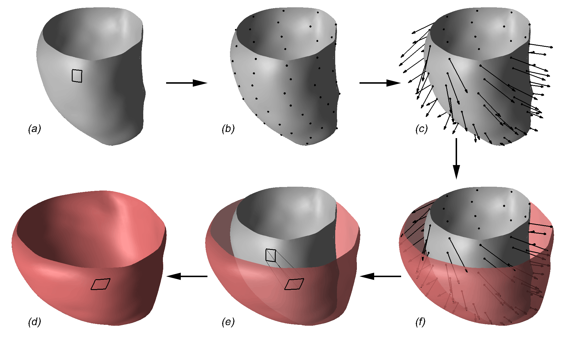


Biventricular Deformation Recovery from Cine MRI

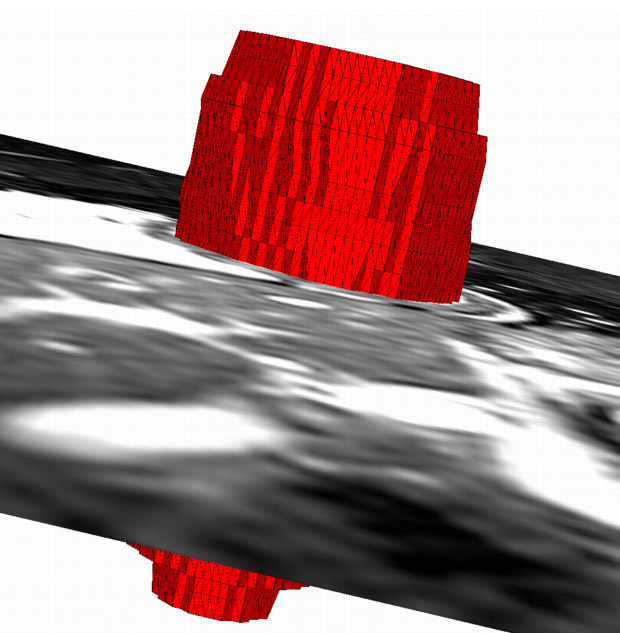
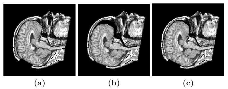
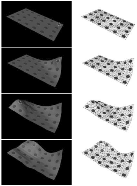
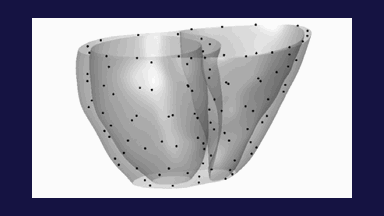
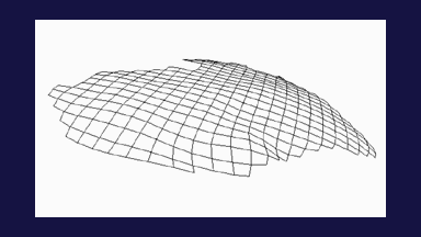
We have developed a method for biventricular myocardial deformation recovery from cine MRI. The method is based on a deformable model that is nearly incompressible, a desirable property since the myocardium has been shown to be nearly incompressible. The model uses a representation that allows for deformation modeling of an arbitrary topology with a relatively small number of parameters, which is suitable for representing the motion of the multi-chamber structure of the heart. The myocardium needs to be segmented in an initial frame after which the method automatically determines the tissue deformation everywhere in the myocardium throughout the cardiac cycle. Fig. 1 demonstrates the types of deformation that the model can generate.
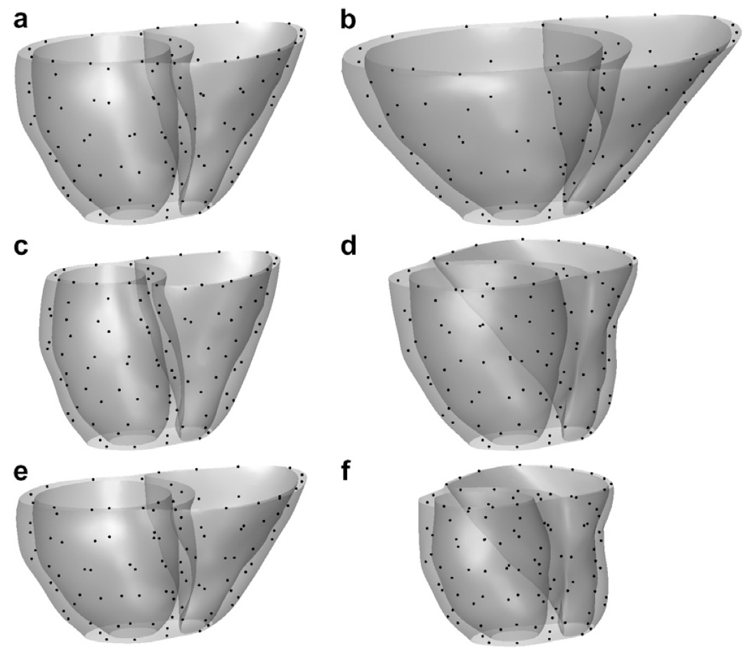
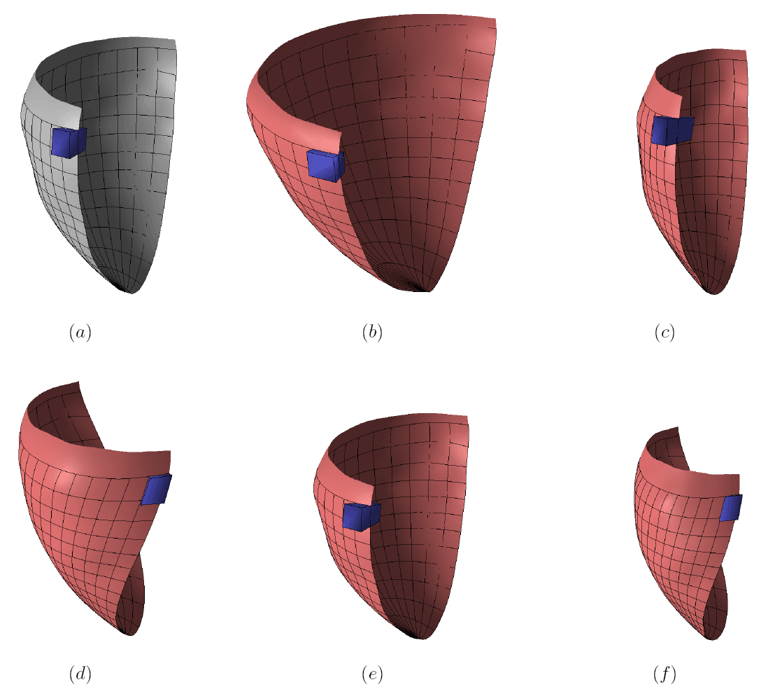
Figure 1: The endocardial and epicardial surfaces of the biventricular model at end diastole (ED) of a healthy volunteer are shown in (a). Artificial transformations are applied to the model to illustrate (b) radial expansion, (c) radial contraction, (d) circumferential twisting, (e) longitudinal shortening, and (f) combined radial contraction, circumferential twisting, and longitudinal shortening, which is a deformation pattern typical for end systole (ES). The black dots represent the model nodes. The figure is from [1] and it is used with permission; Copyright © 2007 Elsevier B. V. All rights reserved.
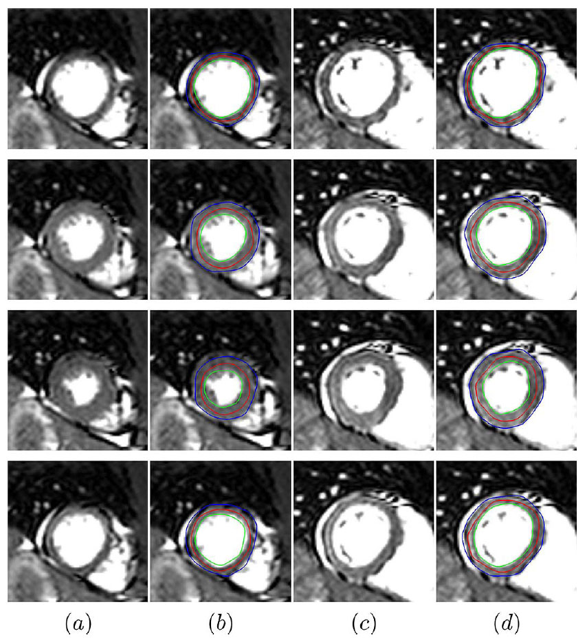
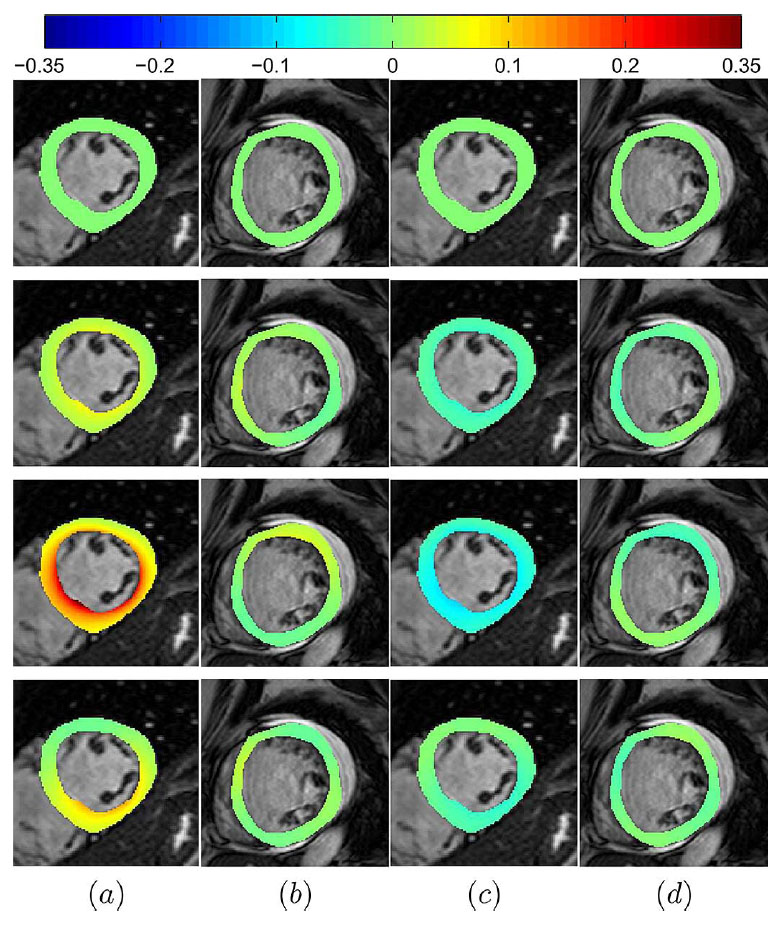
Once the 3D myocardial displacement field is recovered, one can directly compute myocardial strain. Fig. 2 shows radial and circumferential strain of a normal subject in a short axis slice over the cardiac cycle.
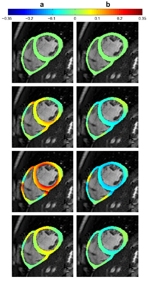
Figure 2: Color-coded (a) radial and (b) circumferential Lagrangian strains for a normal subject are shown in a midventricular slice over the cardiac cycle (first row: ED, third row: ES). Since the deformation is measured relative to ED, the strains in the ED frame are zero. The strains are shown over the ED frame image. The figure is from [1] and it is used with permission; Copyright © 2007 Elsevier B. V. All rights reserved.
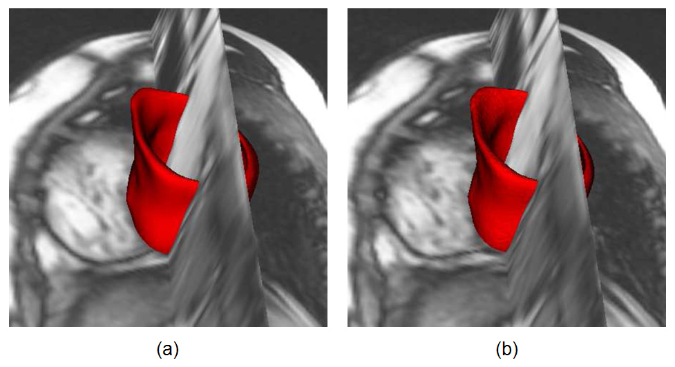
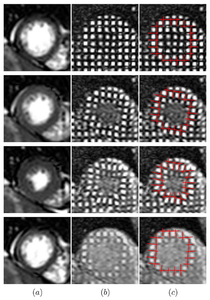
To validate the method, we compared the deformation recovered from a 3-D anatomical cine MRI to the myocardial displacement obtained from corresponding 3-D tagged cine MRI. The average distance between the model and manually determined intersections of perpendicular tag planes was 1.2 pixel. This is illustrated in Fig. 3.
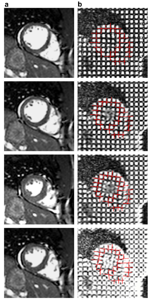
Figure 3: A midventricular short axis slice from the anatomical cine MRI scan is shown in (a) over the cardiac cycle (first row: ED, third row: ES). The corresponding slice in the tagged cine MRI overlaid with virtual tag lines is shown in (b). The virtual tag lines were generated by applying the deformation recovered from the anatomical scan to the manually positioned tag planes at ED. The images are from a healthy volunteer. The figure is from [1] and it is used with permission; Copyright © 2007 Elsevier B. V. All rights reserved.
References:
[1] Bistoquet, A., Oshinski, J., Skrinjar, O., "Myocardial Deformation Recovery from Cine MRI Using a Nearly Incompressible Biventricular Model", Medical Image Analysis, 12(1): 69-85, February 2008. LINK