


Stereo-Guided Volumetric Deformation Recovery

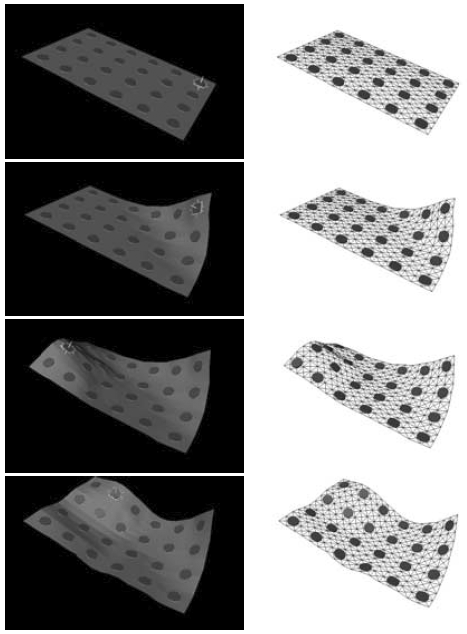
We have developed a framework for soft tissue deformation recovery [1,2,3]. The core of the approach is a volumetric deformable model based on the biomechanics of the material. Often, e.g. in medical applications, the deformation is very complex and dependent on may factors. In such cases, in order to reliably estimate the deformation, the model has to be guided by the available, typically limited, data. We proposed the use of stereo cameras for guidance, where the reconstructed surface is used as a boundary condition for the partial differential equations that define the model. Fig. 1 and 2 illustrate the concept in 2D.
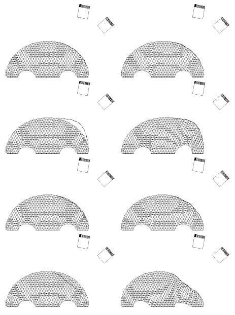
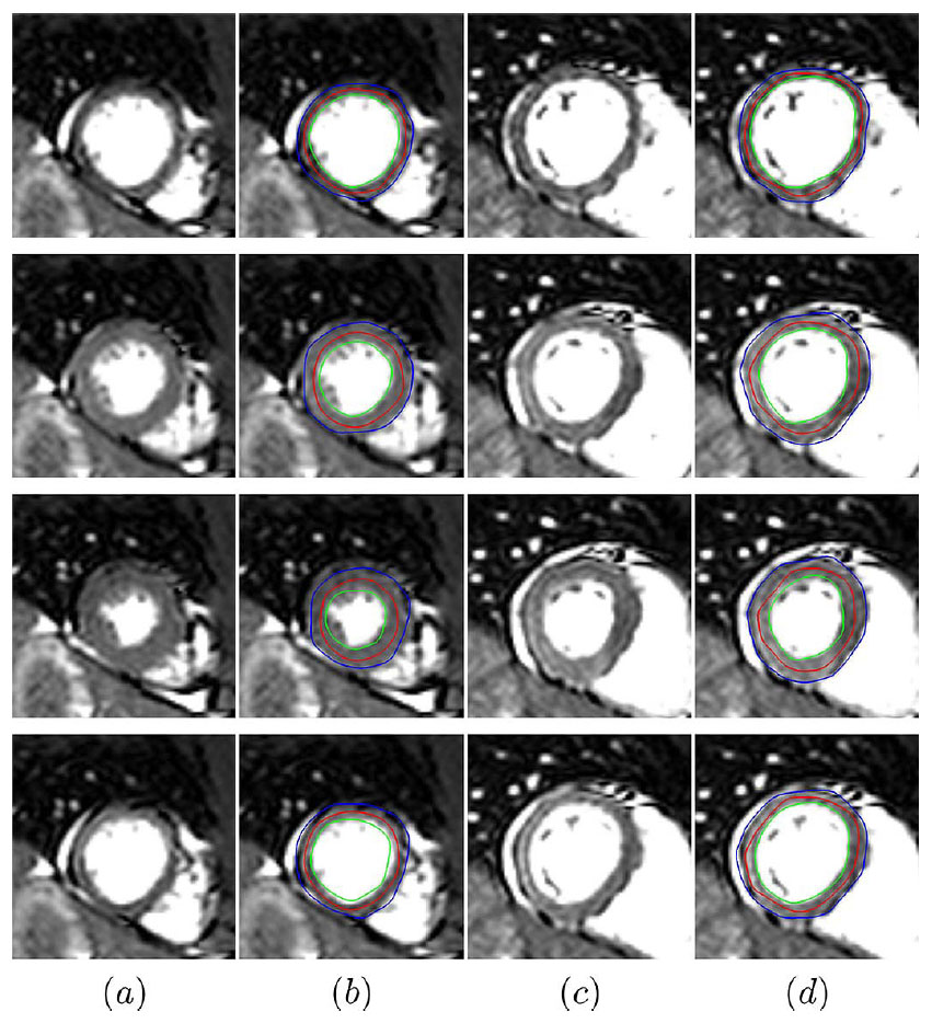
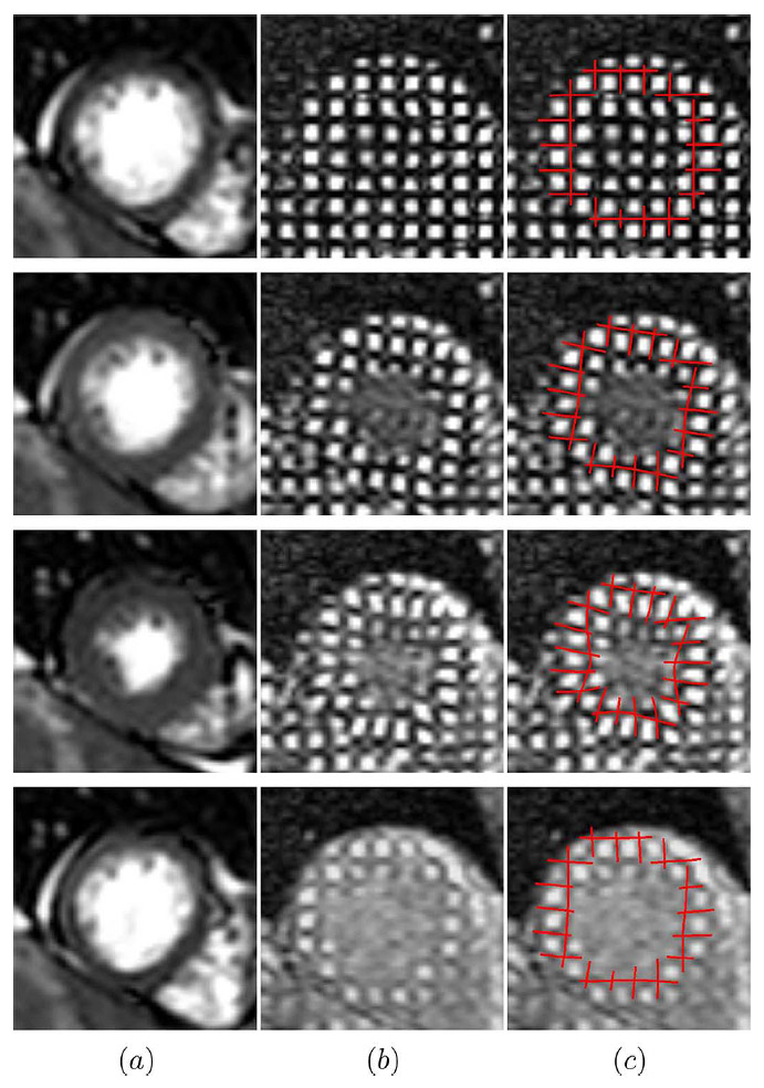
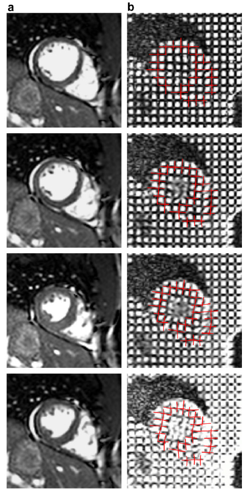
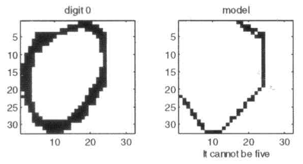
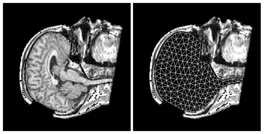
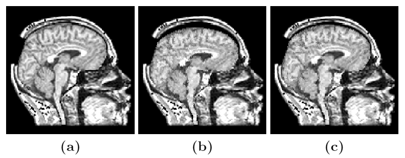
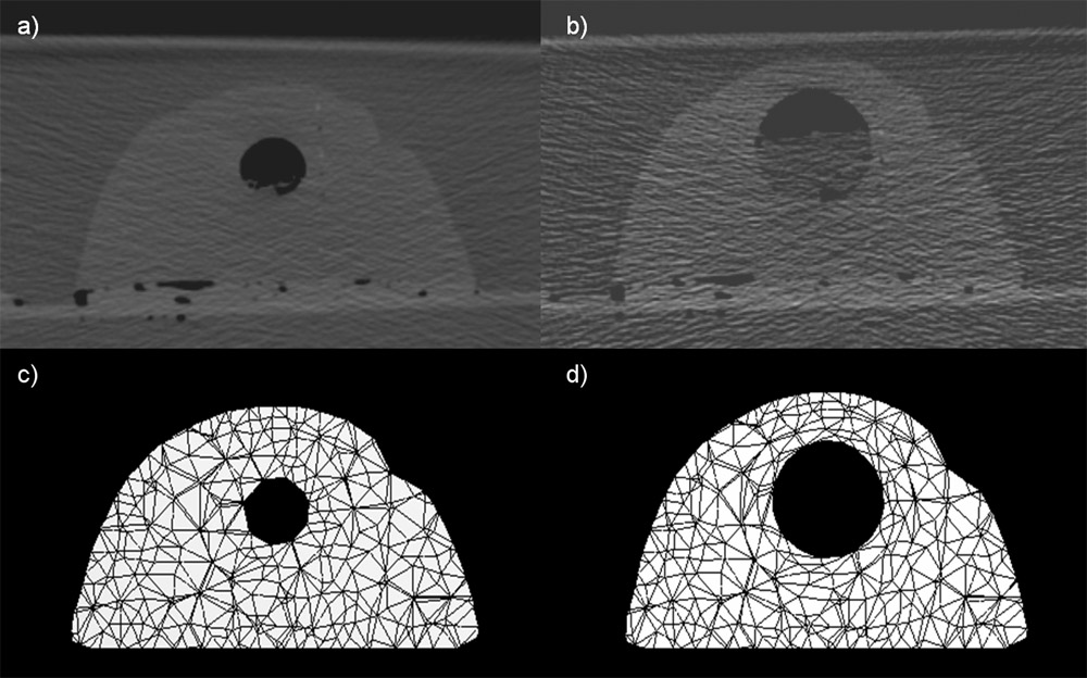
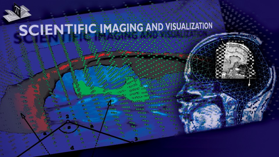
Figure 1: An example of 2D deformation recovery. The left column is a time sequence of the true surface displayed with the undeformed model mesh, while the right column shows the true surface and the updated model mesh using he computed displacement field. A pair of virtual cameras is used for model guidance. The bottom row of model nodes was fixed ("falx"). The figure is from [1] and it is used with permission; Copyright © 2001 IEEE; All rights reserved.
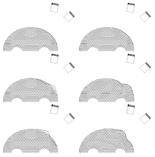
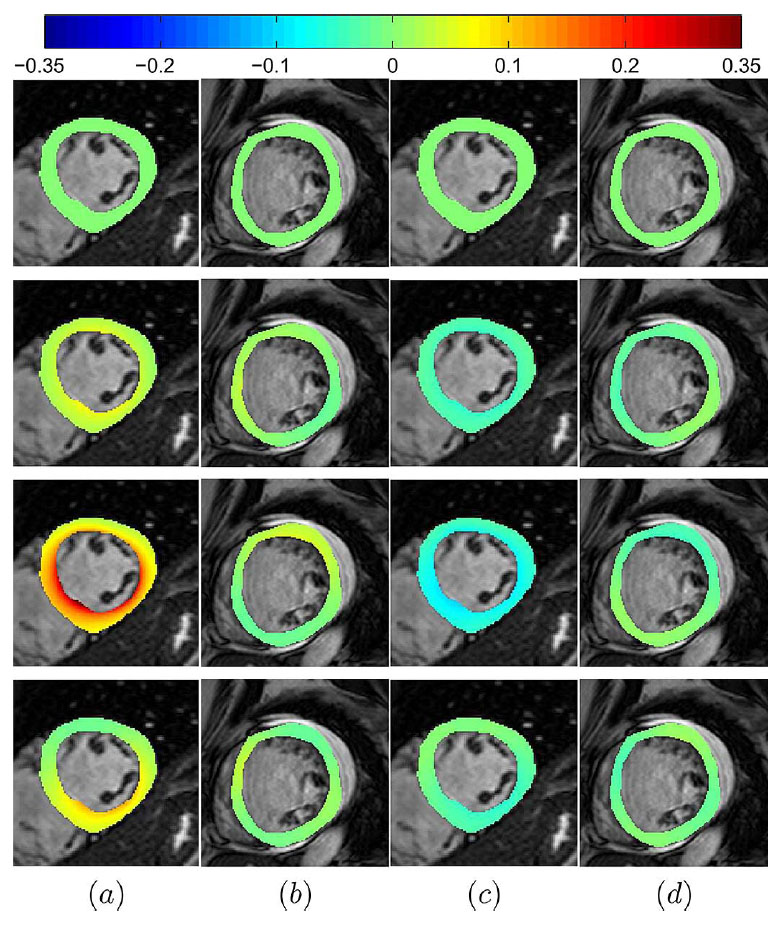
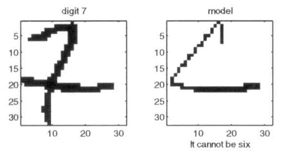
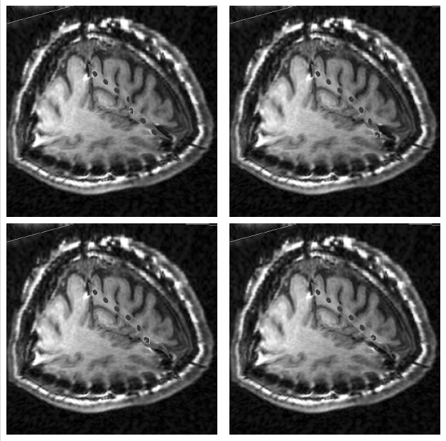
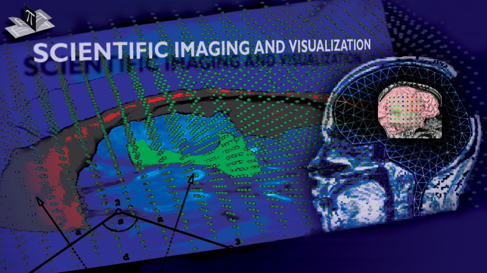
Figure 2: Another example of 2D deformation recovery. The figure is from [1] and it is used with permission; Copyright © 2001 IEEE; All rights reserved.
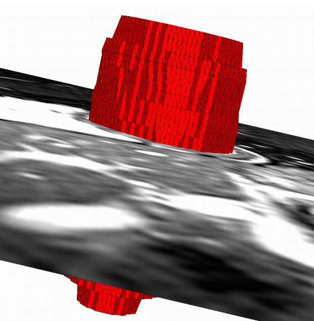
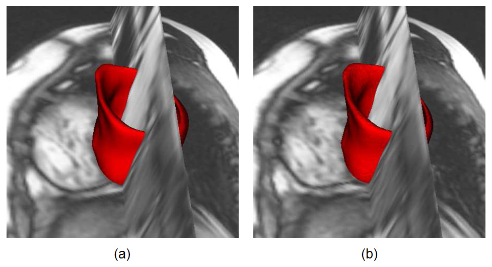
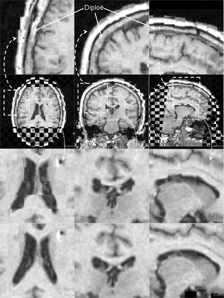
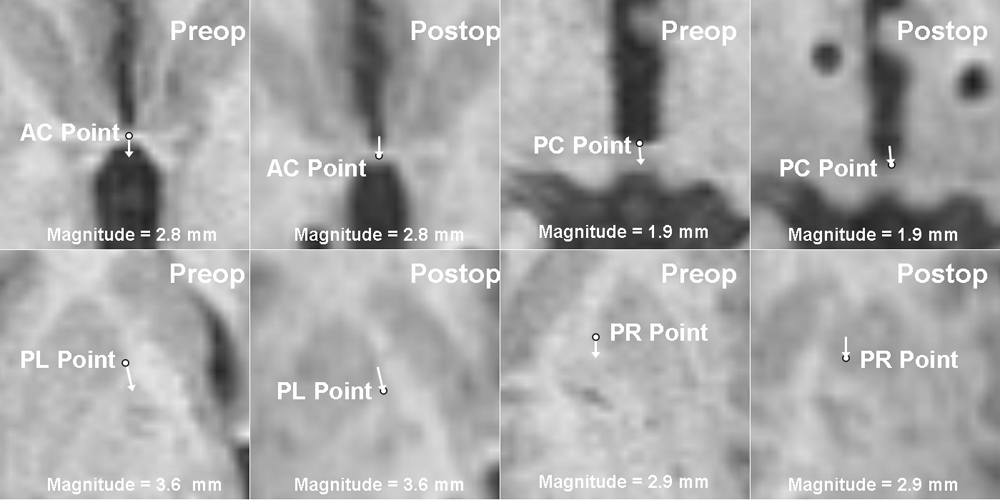
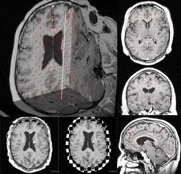
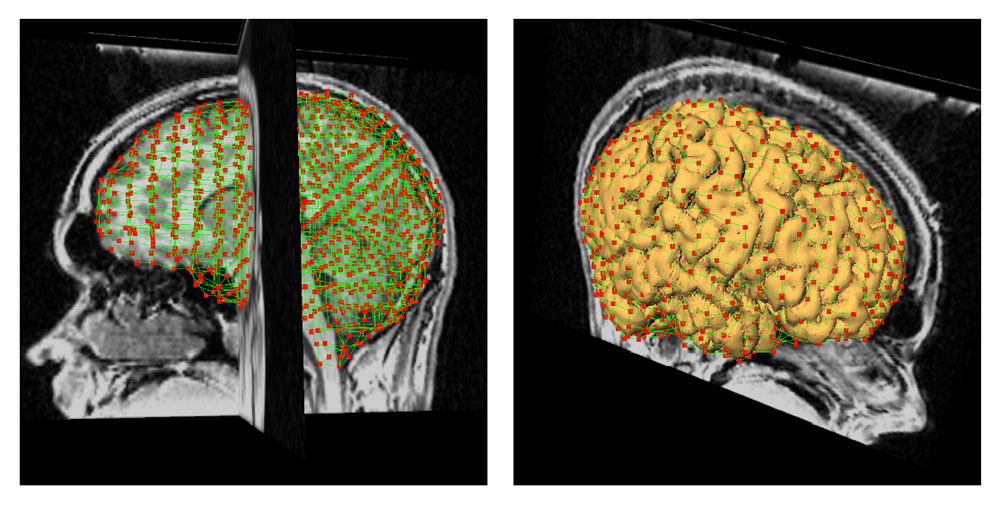
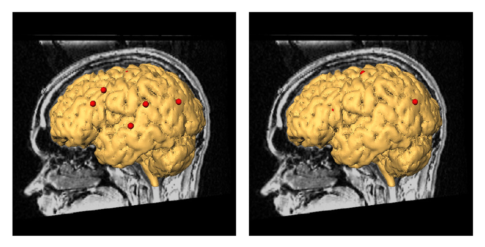
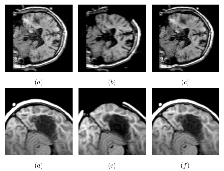
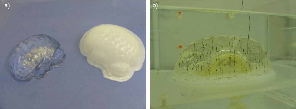
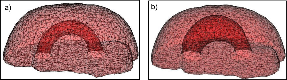
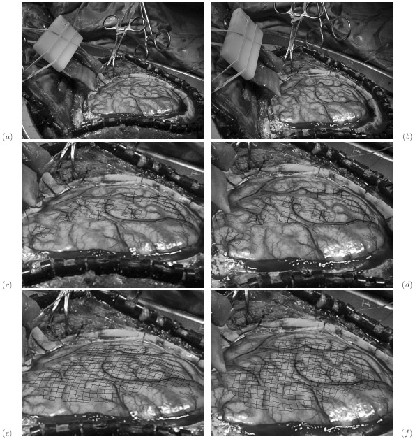
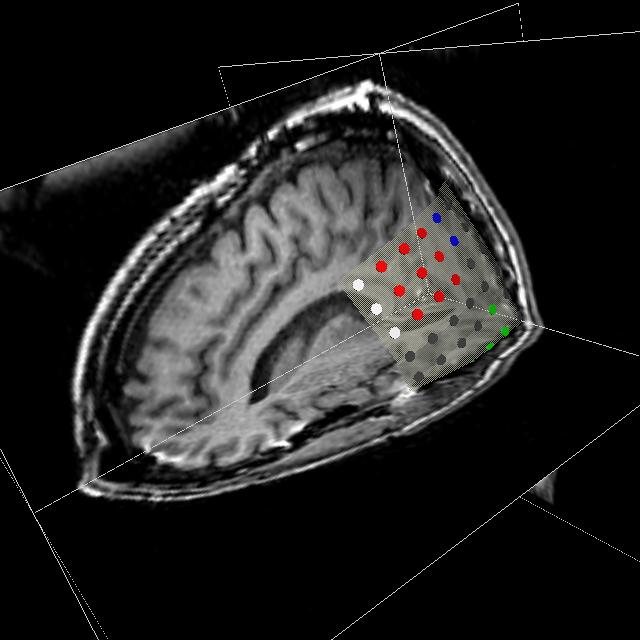
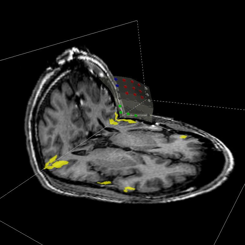
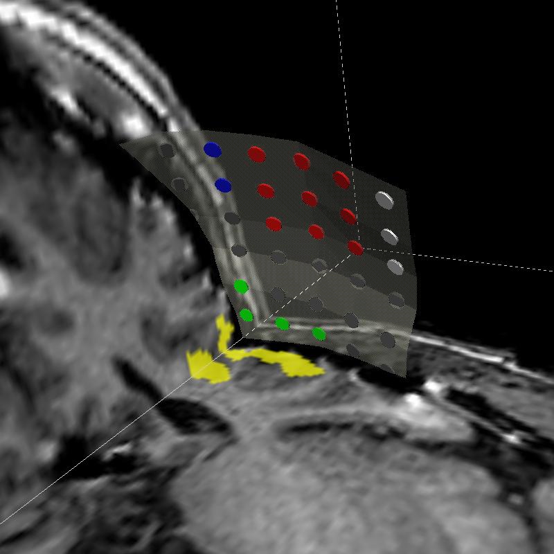
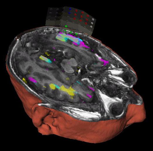
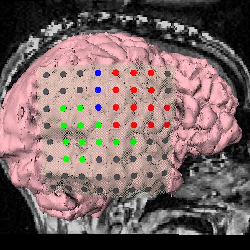
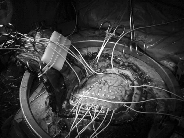
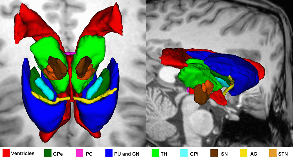
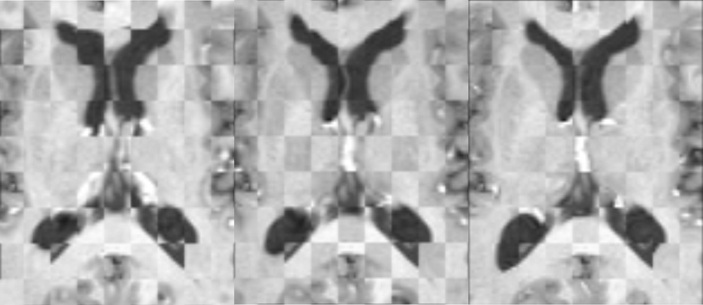
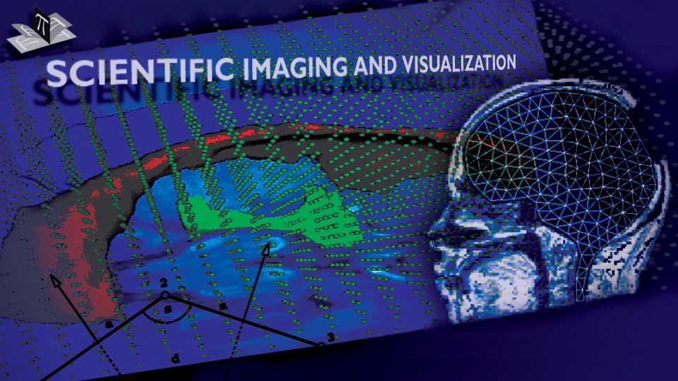
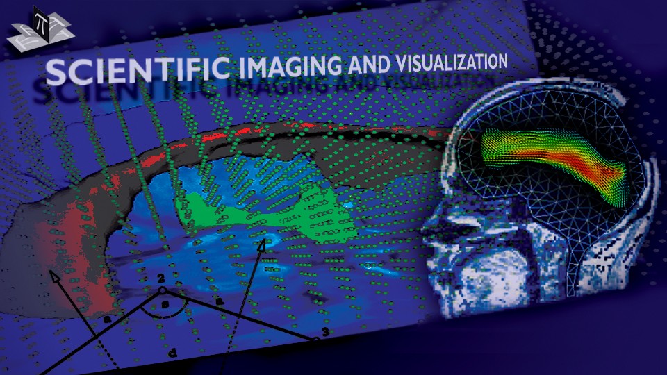
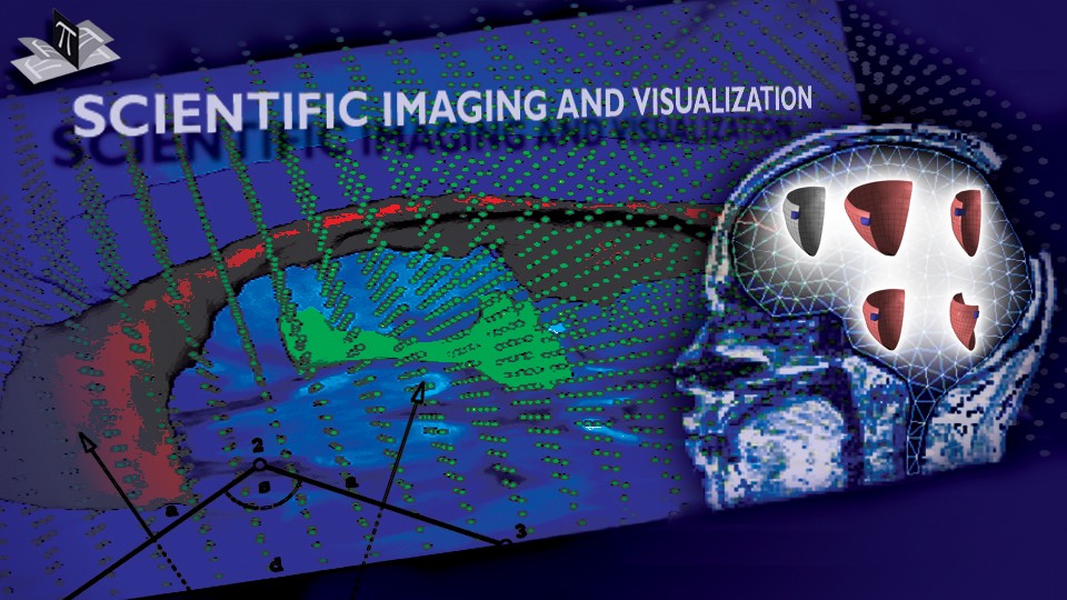
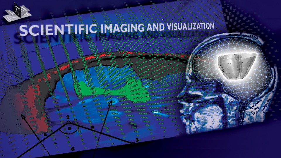
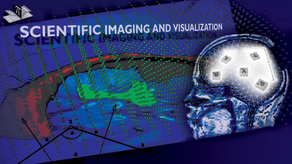
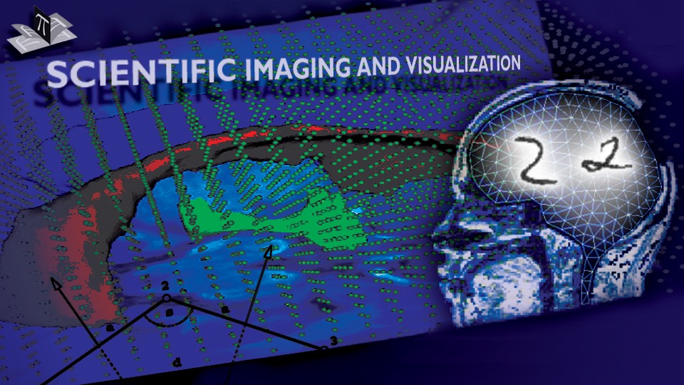
We applied the approach to the problem of brain shift, i.e. brain deformation that occurs during the surgery. Fig. 3 shows stereo views of an exposed brain surface and its reconstructed surface mesh, while Fig. 4 shows 3D simulation results. A partial validation using intraoperative magnetic resonance images suggests that the error introduced by the brain shift can be reduced almost to the image resolution.
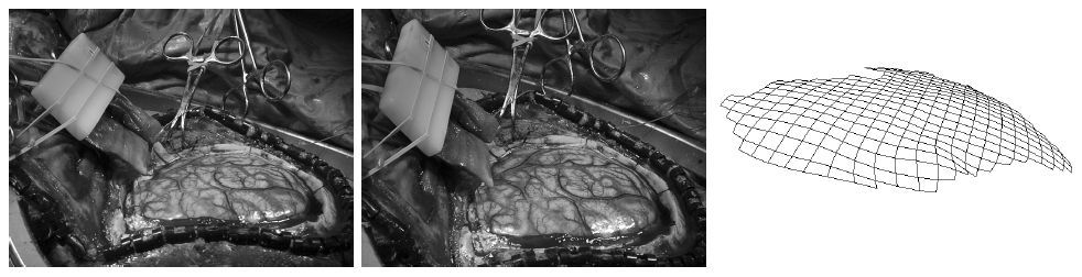
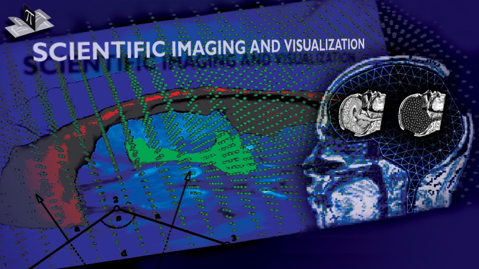
Figure 3: Left and right camera views of an exposed brain surface and a reconstruction of the surface. The figure is from [2] and it is used with permission; Copyright © 2001 Springer Berlin / Heidelberg; ; All rights reserved.
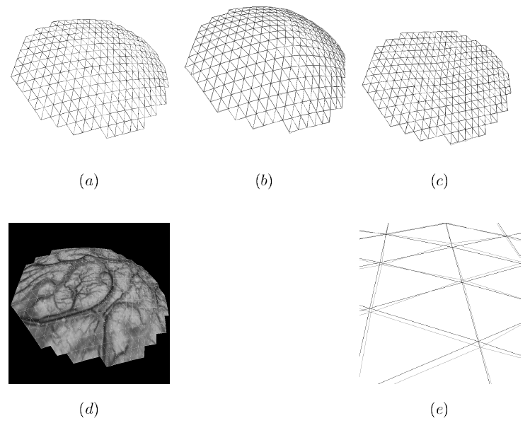
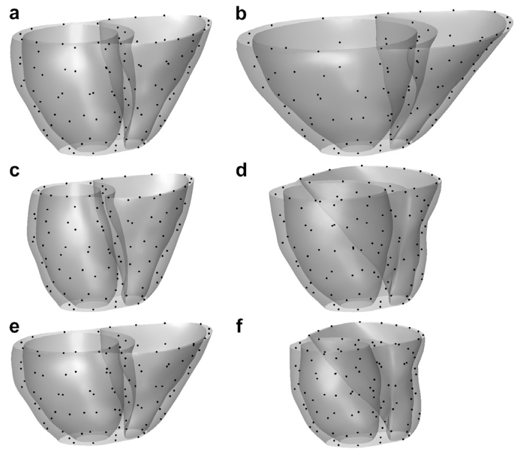
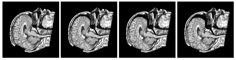
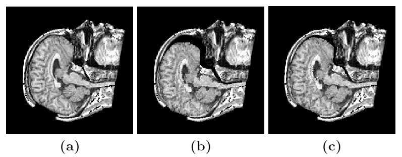
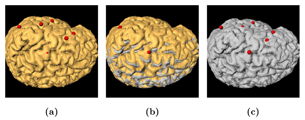
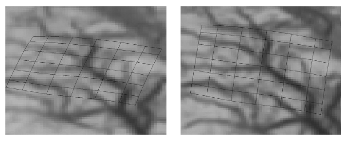
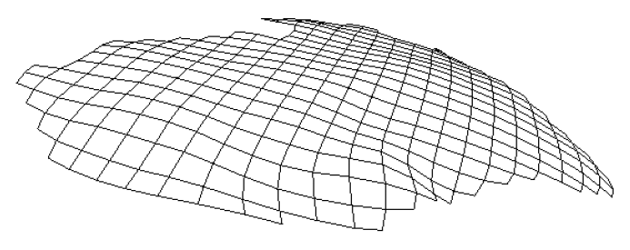
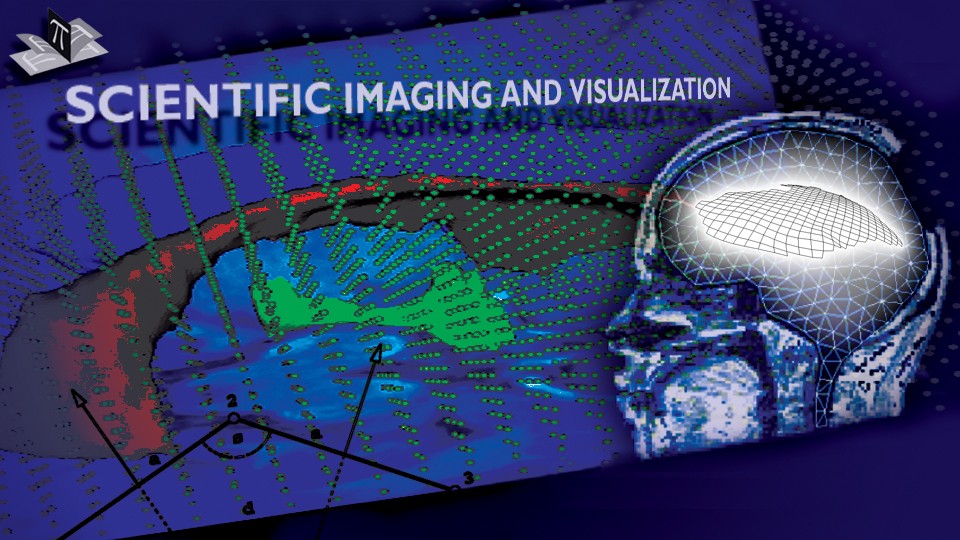
Figure 4: Simulation of an initially bulging and then sinking brain. (d) an exposed brain surface with a texturemap, (a) pre-deformation exposed brain surface mesh, (b) the exposed surface mesh at the peak of bulging (12 mm max displacement relative to the initial surface), (c) the exposed surface mesh at the peak of sinking (28 mm max displacement relative to the peak of bulging), and (e) a part of the zoomed-in true (solid) and recovered (dashed) exposed surface mesh at the peak of sinking (triangle side length approximately 4 mm). The figure is from [3] and it is used with permission; Copyright © 2002 Oskar Skrinjar; All rights reserved.
References:
[1] Skrinjar, O., Nabavi, A., and Duncan, J., "A Stereo-Guided Biomechanical Model for Volumetric Deformation Analysis", IEEE Workshop on Mathematical Methods in Biomedical Image Analysis, Kauai, HI, USA, pp. 95-102, December 2001. LINK
[2] Skrinjar, O., Studholme, C., Nabavi, A., and Duncan, J., "Steps Toward a Stereo-Camera-Guided Biomechanical Model for Brain Shift Compensation", Information Processing in Medical Imaging, Proceedings, Davis, CA, USA, pp. 183-189, June 2001. LINK
[3] Skrinjar, O., "Deformable Models in Image-Guided Neurosurgery", PhD Thesis, Yale University, May 2002. LINK