


Brain Multimodality Registration and Visualization

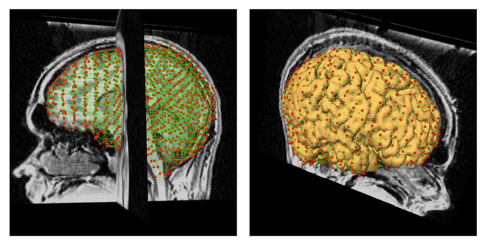
Brain surgery planning can benefit from the use of different imaging modalities and other types of data. We have developed methods for co-registration of different imaging modalities as well as methods for localization of subdural electrodes from MRI head scans and their combined visualization [1]. Figs. 1-5 illustrate combined visualization of anatomical, functional and electro-physiological data.
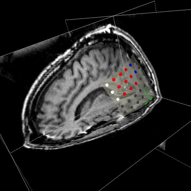
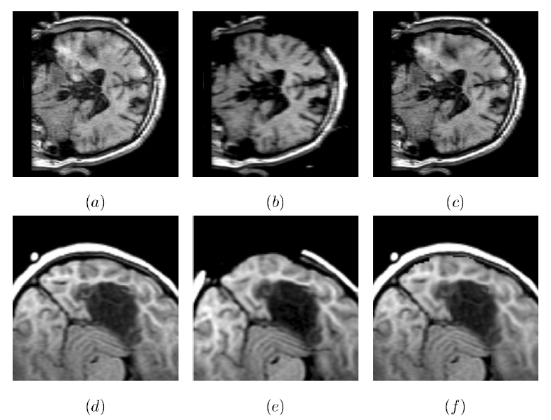
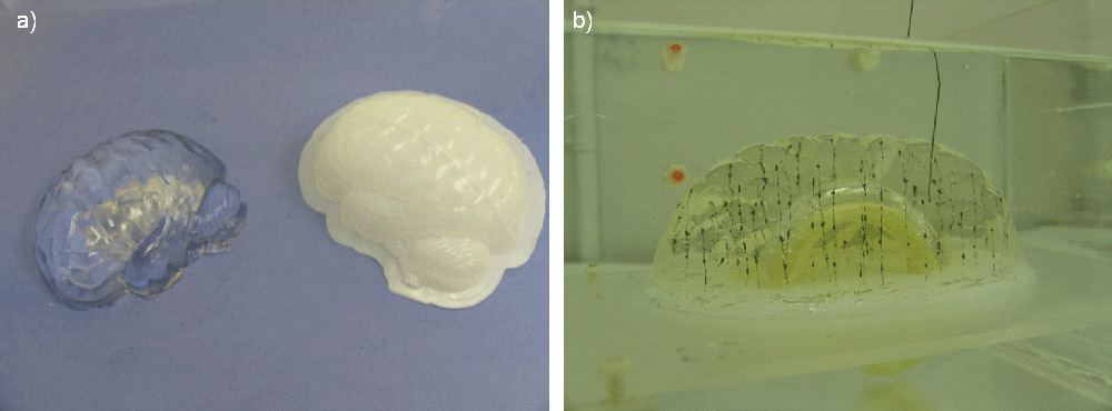
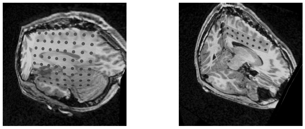
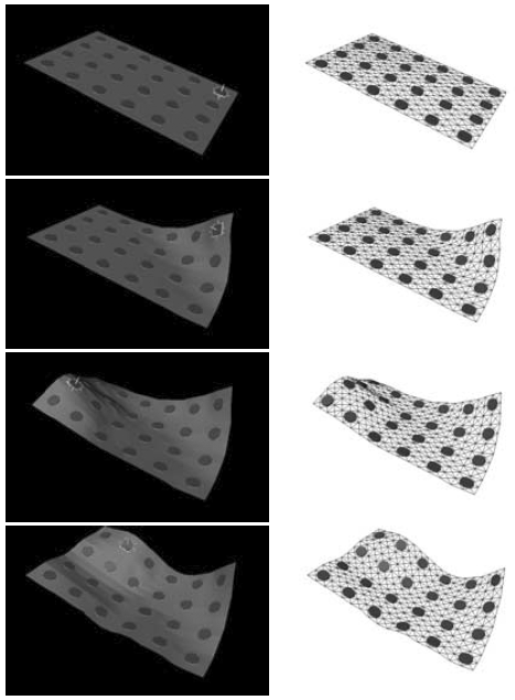
Figure 1: Combined visualization of anatomical and electro-physiological data. An axial, coronal and sagittal slice through the patient's MRI head scan are shown together with a model of an 8 x 8 subdural electrode grid color-coded with electro-physiological recordings. The figure is from [1] and it is used with permission; Copyright © 2002 Oskar Skrinjar; All rights reserved.
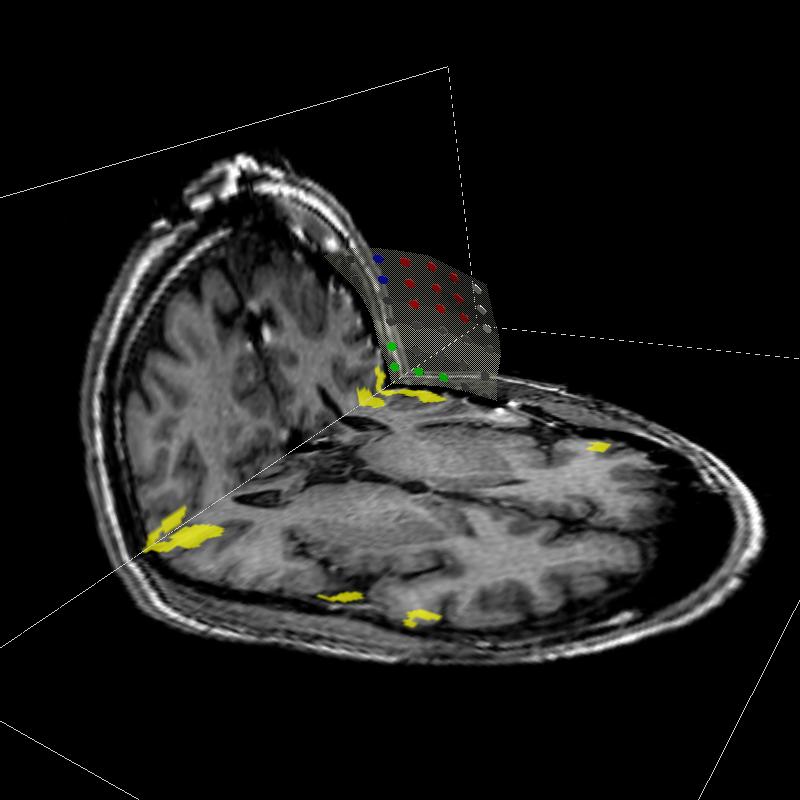
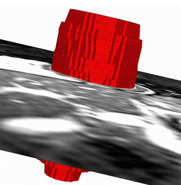
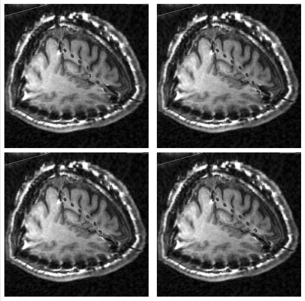
Figure 2: Combined visualization of anatomical, functional, and electro-physiological data. An axial and coronal slice through the patient's MRI head scan are shown together with a model of an 8 x 8 subdural electrode grid color-coded with electro-physiological recordings. The yellow regions are from co-registered fMRI scan. The figure is from [1] and it is used with permission; Copyright © 2002 Oskar Skrinjar; All rights reserved.
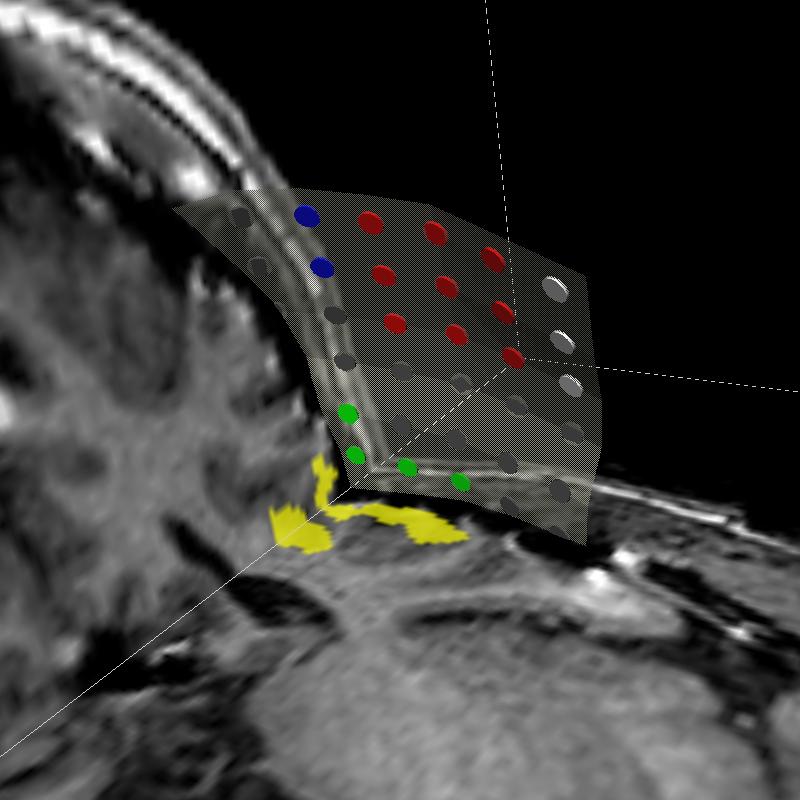
Figure 3: A zoomed-in version of Fig. 2. The figure is from [1] and it is used with permission; Copyright © 2002 Oskar Skrinjar; All rights reserved.
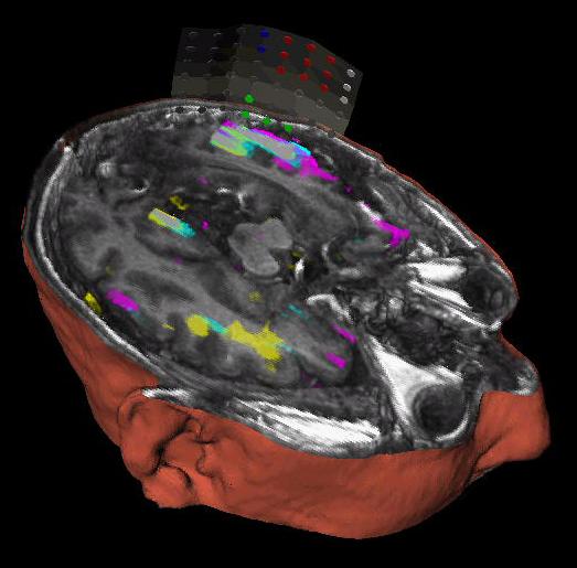
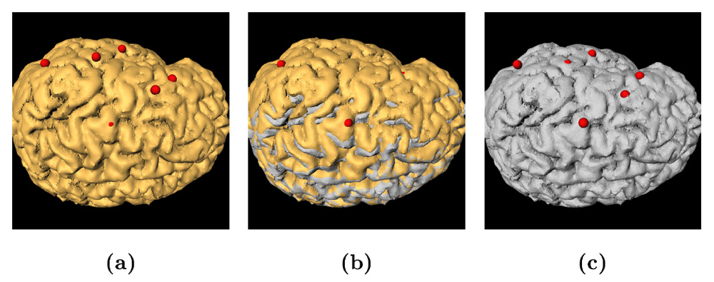
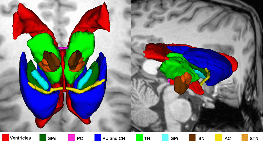
Figure 4: Combined visualization of anatomical, functional, and electro-physiological data. The MRI head scan (gray image) and fMRI scan (colored regions) are volume rendered and clipped with an oblique clip plane, while the outer head surface is surface rendered. In addition, a model of an 8 x 8 subdural electrode grid color-coded with electro-physiological recordings is shown on the brain surface. The figure is from [1] and it is used with permission; Copyright © 2002 Oskar Skrinjar; All rights reserved.
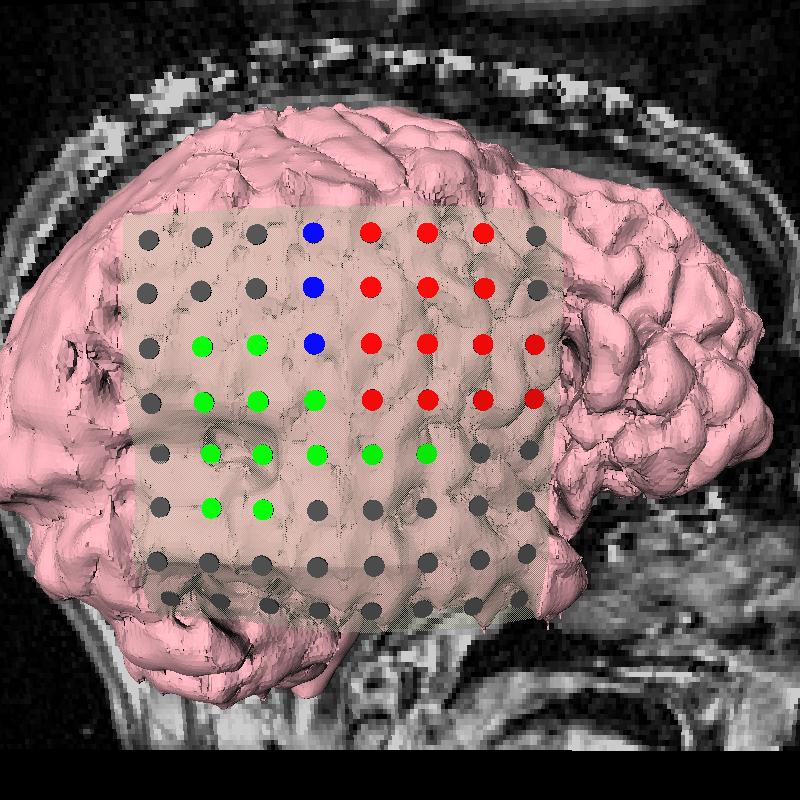
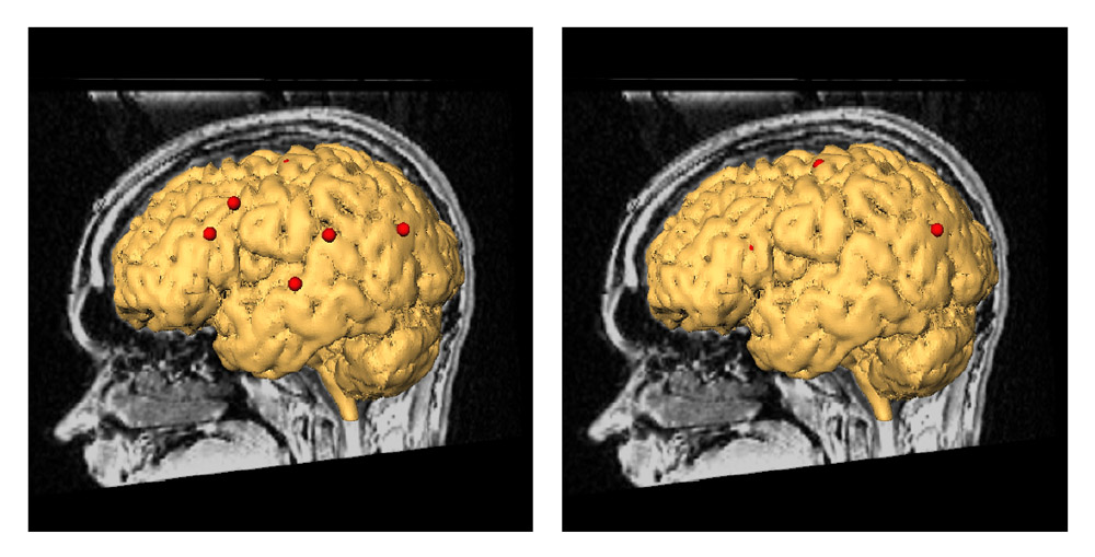
Figure 5: Combined visualization of anatomical and electro-physiological data. A sagittal slice through the MRI head scan is shown together with surface rendered brain surface and a model of an 8 x 8 subdural electrode grid color-coded with electro-physiological recordings. The figure is from [1] and it is used with permission; Copyright © 2002 Oskar Skrinjar; All rights reserved.
References:
[1] Skrinjar, O., "Deformable Models in Image-Guided Neurosurgery", PhD Thesis, Yale University, May 2002. LINK